Sponsored by CareCredit
Lumps on Dogs: Types and What They Mean
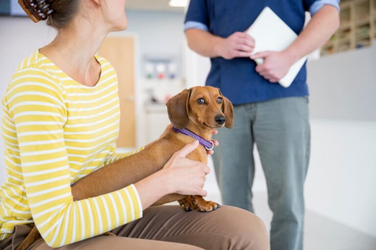
Finding a lump or bump on your dog can trigger a wave of fear and anxiety, especially if it suddenly sprouts out of nowhere. More often than not, our minds immediately jump to the worst case scenario: my dog has cancer. Fortunately, less than half of lumps on dogs are cancerous (malignant) and most are treatable. In fact, lumps on or below the skin are the most common masses reported in dogs, representing roughly one-third of all tumors (both non-cancerous and cancerous).
Most canine lumps look or feel similar regardless of whether they are cancerous. If your dog has a new lump, it’s crucial to have your veterinarian check it to know for sure. While a quick Google search may be tempting, it’s easy for pet parents to misdiagnose a lump and then delay the care their pet actually needs. Early intervention can prevent unnecessary discomfort and potentially save your dog from serious, life-threatening consequences.
Let’s explore some common types of lumps and bumps on dogs, including what they look like and what they mean for a dog’s health and longevity.
First Things First: Don’t Panic
Lumps and bumps on dogs pop up for a variety of reasons, many of which are of little threat to your dog’s life. The bump could be an abscess from a bite, an inflamed hair follicle, or a non-cancerous (benign) fatty growth. In other cases, it could be a more serious condition, like cancer.
Finding a lump on a dog does not necessarily mean the worst, but it is important for a veterinarian to evaluate it so they can make a proper diagnosis and initiate a treatment plan if necessary.
In most cases, you can schedule a vet appointment at your earliest convenience, such as your next day off work. However, if the canine lump or bump is hot to the touch, growing quickly, producing pus or discharge, actively bleeding, or if your dog is in pain, you should take them to an emergency veterinary hospital for more urgent care.
13 Types of Lumps on Dogs

Hard immovable lumps on dogs or sudden lumps on dogs tend to be more worrisome than soft moveable lumps or slow-growing lumps. But that’s not always the case, as many different types of lumps can have a similar appearance.
Lumps on dogs can occur either on the skin surface (cutaneous) or beneath it (subcutaneous).
Common lumps on a dog’s skin (cutaneous masses) include:
- Mast cell tumors
- Histiocytomas
- Perianal gland adenomas
- Sebaceous gland adenomas
- Melanomas
- Squamous cell carcinomas
- Warts
- Hives
Common lumps under a dog’s skin (subcutaneous masses) include:
- Lipomas (tumors of fatty tissue)
- Abscesses
- Soft tissue sarcomas
- Peripheral nerve sheath tumors
- Enlarged lymph nodes
What follows is a more detailed description of each of these types of lumps on dogs:
Mast Cell Tumors
Mast cell tumors are the most common cancerous (malignant) lumps found on dogs. This type of dog skin cancer is usually on the surface of the skin but can be below the skin (subcutaneous) as well.
These masses are usually red, raised, firm, and often form a wound that will not heal. A mast cell tumor typically appears as a hard lump on a dog’s skin that is bleeding, although this can vary greatly.
This cancerous lump on dog skin tends to be aggressive and spread quickly, so it’s important to have it surgically removed as soon as it gets diagnosed. Complete surgical removal is curative as long as the cancer has not yet spread to other parts of the body. In some cases, your dog may need follow-up radiation therapy or chemotherapy.
Histiocytomas
Histiocytomas are non-harmful (benign) skin growths that are most common in young dogs, but they can occur in dogs of any age. Histiocytomas are red, raised, hairless, button-like growths, typically the diameter of a nickel or quarter. They commonly occur as lumps or bumps on a dog’s head or limbs.
Since this red lump on dog skin can appear similar to a mast cell tumor, you should have your veterinarian test it to be certain. Histiocytomas typically regress on their own without any treatment.
Perianal Gland Adenoma
Perianal gland adenomas are common skin tumors of dogs that arise from the glands around the hairless skin of the anus. These are slow growing, benign lumps that occur mostly in non-neutered (intact) male dogs, although they have been reported in spayed females as well.
While these lumps do not spread to other parts of the body, they are locally invasive, meaning they disrupt surrounding tissues, and are prone to infection, so you should not ignore them. The treatment of choice in intact male dogs is castration and tumor removal. Small tumors frequently regress after neutering, and may not require surgical removal. Treated pets typically go on to live long, healthy lives.
Sebaceous Gland Adenomas
Sebaceous gland adenomas are non-cancerous growths that protrude from the surface of the skin. These are usually hairless, firm, small lumps on dogs that occur mostly on the head, neck, back, eyelids, and limbs. These dog cysts can burst open, become irritated, infected, or a combination thereof, but usually they are not problematic.
Your veterinarian may recommend removal if sebaceous gland adenomas are troublesome to your dog. In most cases, no treatment is necessary.
Melanomas
Melanomas are tumors of the cells that produce pigment in animal skin (melanocytes). A melanoma is usually a black lump on a dog, but they are sometimes red. These are hard immovable lumps on dogs. Melanomas most often occur in the oral cavity or on the toes, but these lumps can occur on haired skin as well.
While melanomas on haired skin are usually benign, melanomas found in the mouth or on the toes are usually cancerous tumors in dogs. Malignant melanomas are very aggressive and quickly spread to other parts of the body. Surgery is necessary for treatment of melanomas. Some cases will require chemotherapy or radiation therapy in addition to surgery. The sooner your veterinarian identifies and treats a melanoma in your dog, the better the chances for survival.
Squamous Cell Carcinomas
Squamous cell carcinomas are a common cancerous growth of skin cells in dogs. These lumps can occur anywhere on a dog’s body. This includes lumps on a dog’s legs, abdomen, thorax, toes, paw pads, ears, mouth, or nose. Frequent exposure to UV light is a known cause for developing this type of dog skin cancer. It is more often seen in dogs with light coats or parts of the dog with little coat coverage, like the belly.
Squamous cell carcinomas can appear many different ways. The usual appearance is a single red lump on a dog’s skin. Sometimes they develop as a small area of irritated, red, or ulcerated skin, while other times, they develop as plaques or crusts on a dog’s skin. Carcinomas of the toe or nail bed tend to appear red, irritated, and ulcerated, and are usually quite painful. Dogs may even lose nails on the affected toes.
Treatment of squamous cell carcinomas in dogs is surgery, especially if it is affecting the toe, as it tends to be more likely to spread from that location. If the lump gets removed before it spreads, dogs have a great prognosis and chance of survival.
Warts
Warts (papillomas) are benign lumps on dog skin caused by canine papillomaviruses. Dog warts are usually small and light-colored with a rough, jagged appearance. These lumps are mostly found in or around the mouth, on the feet, or on the eyelids, but they can grow anywhere on the body. Young dogs less than 2 years old are the most commonly affected.
Dog warts often disappear spontaneously as the dog develops immunity against it. However, some dogs may need to have their warts surgically removed if the warts become irritated, infected, cause pain, or fail to regress on their own.
Hives
Hives in dogs are similar to hives in humans. They appear suddenly as red, raised, circular bumps on the surface of the skin and can occur anywhere on a dog’s body, including in their mouth. Hives in dogs can vary from a few millimeters to several centimeters in size. If hives become large enough, they can blend together, or coalesce.
Direct contact with an allergic substance, such as an insect bite, food, pollen, mold, vaccinations, or medications, can cause hives. Typically, hives are self-limiting and resolve after removal of the allergic substance. Nevertheless, allergic reactions can be severe, and potentially life threatening, so you should notify your veterinarian immediately if you notice hives on your dog.
Lipomas
Lipomas (tumors of fatty tissue) are the most common benign tumor of dogs. These fatty lumps on dogs feel soft or squishy and are usually freely moveable beneath the skin. This means that they are not fixed in place or attached to underlying tissues. Fatty tumors commonly show up as a lump on a dog’s chest or abdomen but can occur anywhere on the body. Some lipomas will hardly grow after initially developing, while others seem to grow relatively quickly.
Although lipomas are non-cancerous, they can still be problematic if they develop in places that impair a dog’s ability to walk or lie down. If a lipoma is growing quickly or in a worrisome location, a veterinarian will typically recommend surgical removal.
Abscesses
Abscesses in dogs are pockets of pus underneath the skin. These are usually soft lumps on dogs that are warm to touch and painful, and occur secondary to a bite wound or skin injury. Abscesses in dogs are often just below the skin, can be large or small, and sometimes rupture and drain a foul smelling fluid. They can occur anywhere on the body. Abscesses are also seen on the muzzle of dogs, usually underneath the eye, secondary to dental disease.
Dogs with abscesses will need veterinary care to have the abscess drained and flushed out. They will need antibiotic therapy as well as pain medications. Abscesses in dogs are usually so painful that many pets will need some level of sedation. This allows the veterinarian to treat the issue properly and spare canine patients from more pain or discomfort.
Soft Tissue Sarcomas
Soft tissue sarcomas are a category of cancerous tumors, particularly those arising from the connective muscle or nervous tissues in dogs. Since these tissues are present throughout the entire body, these tumors can develop anywhere. They occur most often on the legs, chest, or abdomen of affected dogs, and are more common in middle aged to older dogs.
Soft tissue sarcomas are typically hard immovable lumps on dogs found beneath healthy skin. In most cases (but not all), these cancerous tumors in dogs do not typically spread but will grow into and disrupt surrounding tissues.
Surgical excision is the best treatment for soft tissue sarcomas in dogs. The surgeon must take wide margins to avoid leaving any of the cancer cells behind, so it is ideal to remove these tumors while they are still small. In some cases, your dog may need followup chemotherapy or radiation therapy. After successful surgical removal, most dogs will live full lifespans.
Peripheral Nerve Sheath Tumors
Peripheral nerve sheath tumors are a type of soft tissue sarcoma. These lumps grow from the nerve cells and can occur anywhere in a dog’s body. They feel like hard immovable lumps on dogs under the skin when they grow close to the surface. These lumps are usually not painful when touched, but some dogs will bite or chew at them due to nerve irritation.
Peripheral nerve sheath tumors do not commonly spread but are locally invasive. Treatment of choice is surgical removal of the tumor, although recurrence is common. In some cases, dogs will need to have the affected limbs amputated and/or undergo radiation therapy. Unfortunately, most dogs with peripheral nerve sheath tumors may only live up to one year, despite appropriate treatment.
Enlarged Lymph Node
Lymph node enlargement in dogs occurs for many reasons, such as infection, inflammation, or cancer. Enlarged lymph nodes are firm, moveable lumps felt underneath healthy skin of dogs. They are usually detected under the chin, on the neck, the front of the shoulder, or the back of the rear leg. Enlargement of a single lymph node is not as worrisome as enlargement of multiple lymph nodes, which is often indicative of systemic disease or cancer.
Treatment of lymph node enlargement in dogs depends on the underlying cause. Your veterinarian may recommend various medications, chemotherapy, or even surgery based on the cause of the lymph node enlargement. Depending on the cause, the long-term prognosis can vary considerably.
Diagnosing Lumps on Dogs

As mentioned earlier, your veterinarian should check out any new lumps or bumps on your dog. A thorough physical exam can help your veterinarian narrow down a list of possible diagnoses. Additionally, you can help out by providing a detailed history of your pet.
Let your veterinarian know whether the issue developed as a sudden lump on your dog under the skin or on the surface of the skin, if you’ve noticed any changes in your dog’s behavior, and whether the lump has grown or changed since you first noticed it.
Many different types of lumps and bumps on dogs can appear and feel similar, so testing the lump to determine exactly what it is and whether it could be potentially problematic for your pet is always advised. Your veterinarian may recommend bloodwork and/or imaging, such as radiographs (X-rays), based on your dog’s specific case. There are financing options available that can help, such as the CareCredit credit card.* Knowing that you can finance the laboratory and diagnostic services your dog needs can give you peace of mind.
The first recommended test is usually a fine needle aspirate and cytology. This is a quick, non-invasive, and affordable test in which your veterinarian will use a needle to suck out (aspirate) cells from the lump to determine their origin and behavior. The cells are then smeared on a glass slide, stained, and evaluated under the microscope.
In some cases, your veterinarian will recommend biopsy. This is a more invasive procedure in which your veterinarian will take a small chunk of tissue from the lump and send it to a pathologist. Your dog will likely need some form of sedation for this procedure but can usually return home the same day. Biopsy samples are almost always diagnostic and can tell you exactly what is causing your pet’s lump and what treatment options are available.
Treating Canine Lumps

After your veterinarian tests the lump and confirms a diagnosis, they will guide you through treatment options for your dog, if necessary. As discussed, treatment can vary greatly depending on the type of lump your dog has.
Some common forms of treatment for lumps on dogs include:
- Surgical removal
- Amputation
- Draining (for abscesses)
- Medications
- Topical ointments (for skin conditions like hives)
- Chemotherapy (if cancerous)
- Radiation (if cancerous)
Even when a lump is cancerous or malignant, your dog may have a great outcome, especially if you start treatment early and aggressively.
The cost of treatment for lumps on dogs varies based on the diagnosis. However, treatment will always be more affordable if you properly address and care for the lump early on, while it is smaller and less likely to have caused secondary issues, like infection, that will also require treatment. Since the type of treatment and associated costs can vary greatly, it’s wise to be financially prepared. Whether your dog requires surgery, chemotherapy, or medication, CareCredit gives you the flexibility to use your card for your pet’s treatments and procedures at locations in the CareCredit network.*
Remember, as a responsible pet parent, you can help identify a new lump or bump on your dog so your veterinarian can diagnose and treat it. Check your pet frequently so you can spot any abnormal growths and have them tested and treated as soon as possible.
*Subject to credit approval
This information is shared solely for your convenience. Neither Synchrony nor any of its affiliates, including CareCredit, make any representations or warranties regarding the products described, and no endorsement is implied. You are urged to consult with your individual veterinarian with respect to any professional advice presented.


